Research
Sensory processing and cognition
I am interested to understand what cognition means in biological terms and how abstract cognitive variables are implemented by cortical networks such as the prefrontal cortex. Brain circuits are continuously performing neuronal computations allowing organisms to act upon hidden rules of the external world. The capacity to extrapolate outcomes from limited observations, decipher rules from complex situations, and maintain focus on specific external inputs exemplifies the abstract cognitive operations performed by various species. By studying the neuronal activity of model organisms during well controlled behavioral paradigms, we hope to reveal the neuronal correlates underlying the abstract cognitive varibles. To achieve this goal we need to have a good knowledge of "the basics" i.e. how neuronal networks are anatomically arranged and how do they respond to simple sensory and motor information. The following paragraphs develop how I think we should tackle the thorny problem of cognition in modern neuroscience with our current tools. The perspectives shared on this website are personal and may not comprehensively capture the diverse range of positions within the field.
Before diving deeper, it's important to acknowledge that research is a collaborative endeavor, and I want to express gratitude to all the individuals I've had the privilege of working with. My academic journey has been shaped by the guidance of exceptional scientists, and I am continually benefiting from their insights. I was fortunate to be trained to handle the electrophysiological tools in the lab of Pr. Paul Salin working side by side with Dr. Sylvain Crochet. I got the unique opportunity to collaborate with Pr. Carl Petersen in Lausanne with whom we designed a long-term project together with Sylvain Crochet including a goal-directed behavioral task and optogenetic photoinhibition experiments in the head-restricted mouse preparation. We found out together a crucial role of the prefrontal cortex and the hippocampus in a simple sensory detection task. Presently, I am exploring the neuronal mechanisms underlying the computational sensory and abstract properties of the prefrontal cortex in the inspiring scientific environment fostered by Pr. Marie Carlén. It's essential to recognize that every person I've encountered throughout my journey—be they students, PhDs, postdocs, technicians, professors—has played a role, directly or indirectly, in shaping the ideas presented here.
The prefrontal cortex
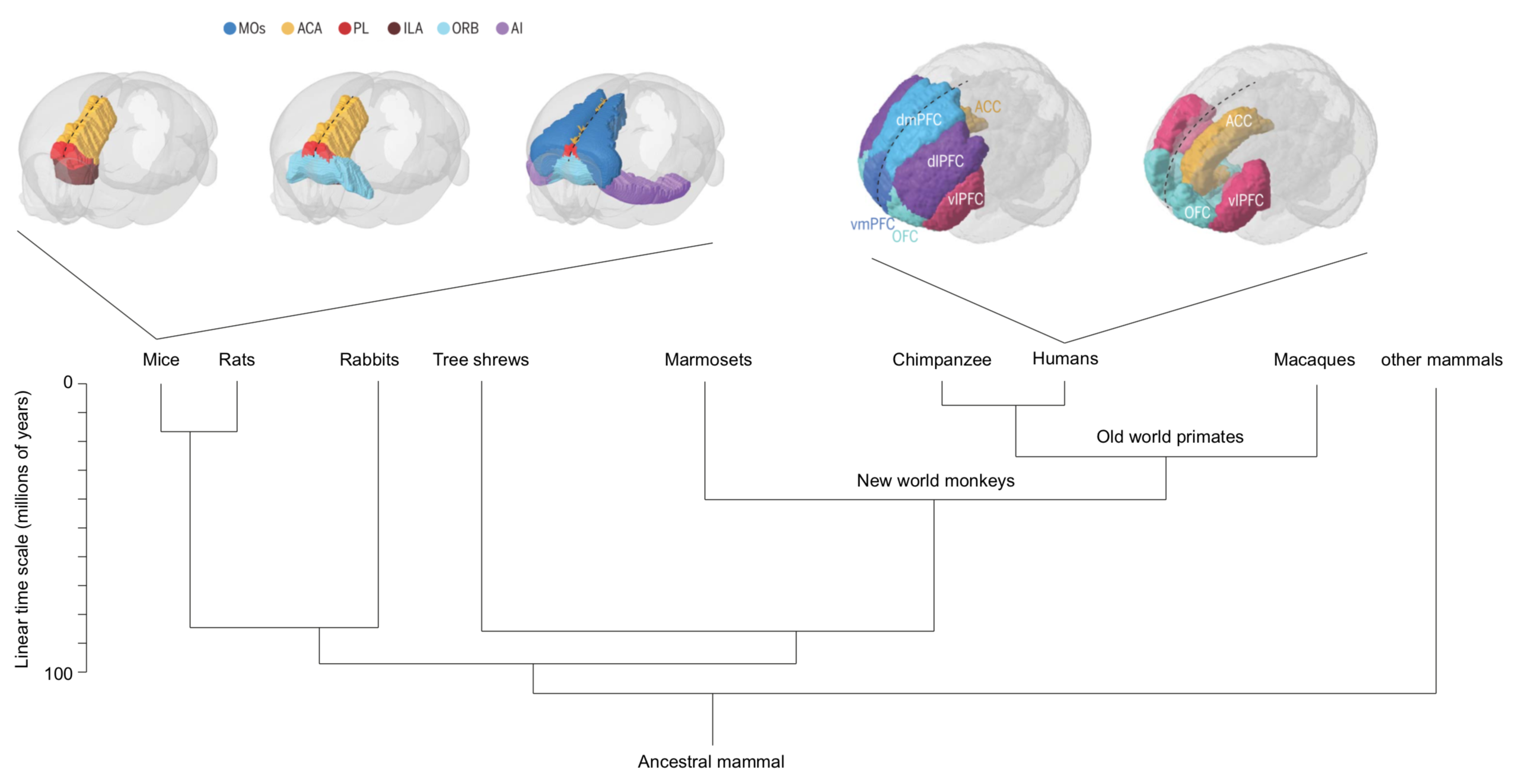
The prefrontal cortex (PFC) is an umbrella term referring to cortical regions located in the front of the brain. The PFC still lacks a conclusive definition, and the structure and function of this brain area across species remain unresolved. The PFC is implicated in perceptual, emotional, social, motivational, and numerous other brain processes and considered to enable flexible behavior. In following, disturbed PFC functioning has been connected to most, if not all, mental disorders, including drug addiction. Present-day preclinical researchers increasingly utilize mice (Mus musculus) as model animals. However, in parallel clinical transfer of pre-clinically identified therapeutics targeting mental disorder (and other brain disorders) is still at large failing. Lack of understanding of the structure and function of the brain hampers the understanding of which findings are transferable between species. Needless to say, deciphering of the structure and function of the PFC is of great importance to medicine. While class-common functions and class-common behaviors have been firmly established, effort must also be put into clarifying dissociations, and perhaps most so between primates and rodents. Ultimately, understanding of what makes the human PFC unique will build on comparative studies in different species.
Cytoarchitecture of the mouse (pre)frontal cortex
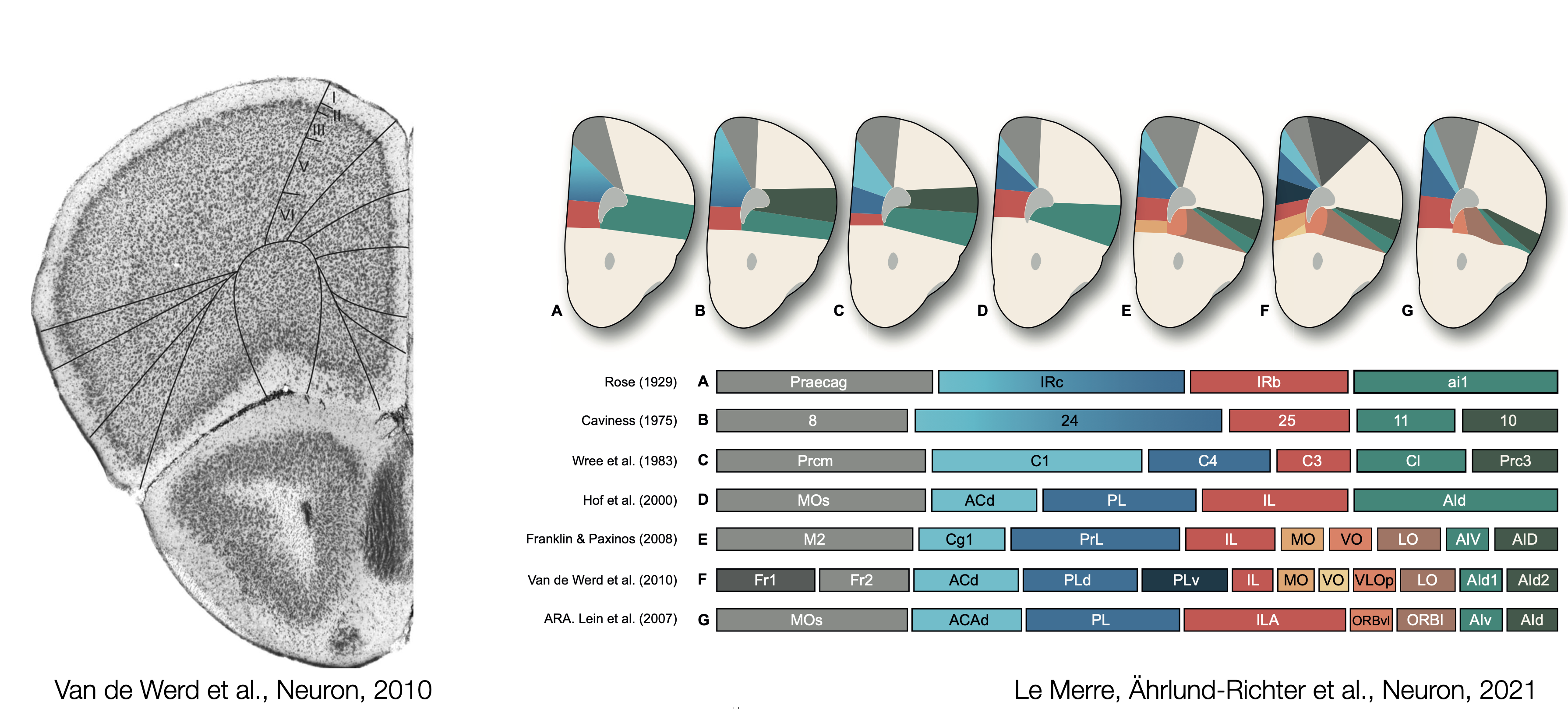
The first maps of the mouse prefrontal cortex come from cytoarchitecture and the seminal work of Korbinian Brodmann where the shape and the size of neurons determine brain regions. Recent atlases divide frontal regions into the Anterior Cingulate areas (ACA; dorsal and ventral), the Prelimbic area (PL), the Infralimbic area (ILA) and Orbital areas (ORB; medial, ventrolateral and lateral). These regions are flanked by the Secondary Motor area (MOs), the Frontal Pole (FRP), Agranular Insula areas (AI; anterior and ventral), the Dorsal Peduncular area (DP) and the Taenia Tecta (TT; dorsal and ventral). The caveat of cytoarchitectural patterns defining regions is the lack of reproducibility and agreement regarding the number and the borders of delineated regions. Over the last century, the names and borders of mouse prefrontal regions have kept changing.
Connectivity of the mouse frontal module
As neuron extend their axons across brain regions, they define the main roads that can be taken to exchange information. To establish how the prefrontal module communicate with the rest of the brain, the main neuronal pathways need to be identified. Viral delivery of fluorescent proteins into genetically targetted neuronal populations has become, over the last two decades, the standard technique to trace afferent and efferent connections of a brain region. The high diversity of mouse lines and the access to a large viral toolbox are the essential components for the prefrontal circuit dissection.
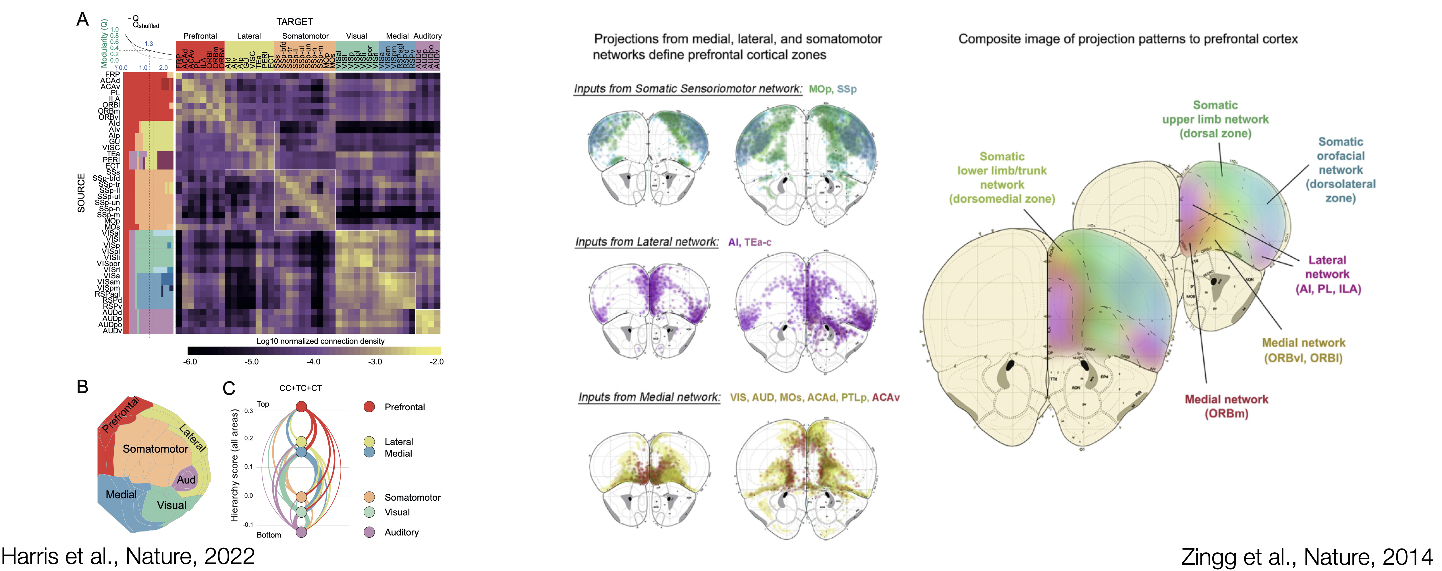
Recent work from the Allen Institute of Science has identified cortical modules based on connectivity patterns. Modules are brain region more interconnected with themselves than with the rest of the brain. The PFC stands as a coherent cortical module encompassing the Anterior Cingulate areas (ACA; dorsal and ventral), the Prelimbic area (PL), the Infralimbic area (ILA), Orbital areas (ORB; medial, ventrolateral and lateral) and the Frontal Pole (FRP). Interestingly, the Secondary Motor area (MOs), and Agranular Insula areas (AI; anterior and ventral) are the last one to jump out of the prefrontal module, potentially indicating that some parts of MOs and AI could be considered more "prefrontal". Taken together, the identified prefrontal module is the cortical module with the most extensive afferent and efferent connectivity with the rest of the brain, forming large brain-wide networks of connected regions.
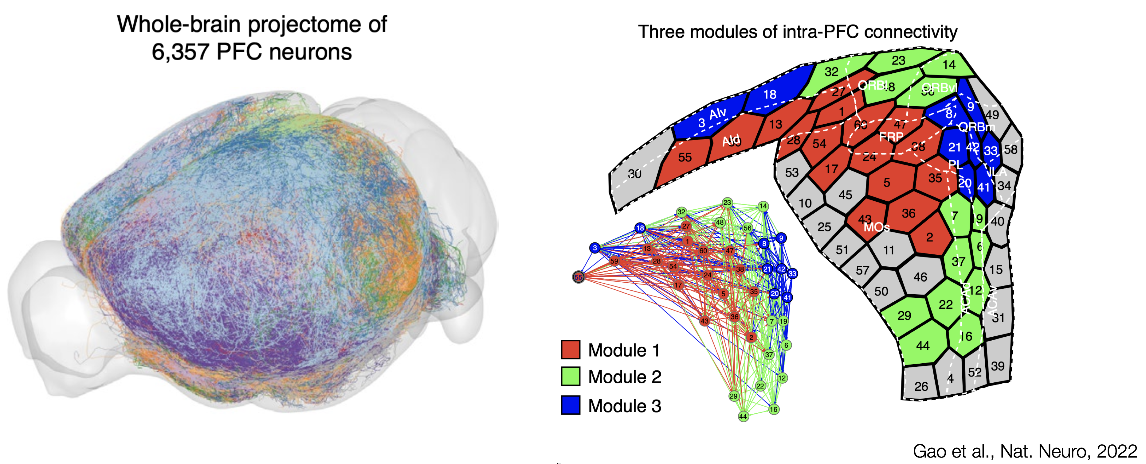
A single-neuron approach, tracing 6,357 single prefrontal neurons axonal projection, has recently been used to delineate interconnected modules within the mouse PFC. From the work of Gao et al., the main three connectivity-defined modules do not abide by cytoarchitectural boundaries. Connectivity eventually orchestrate brain activity by allowing information to flow between brain regions. The lack of agreement between cytoarchitecture and hodology questions the definition of prefrontal regions: What should we take into account to reach a comprehensive definition of the PFC?
Molecular maps of the mouse frontal cortex
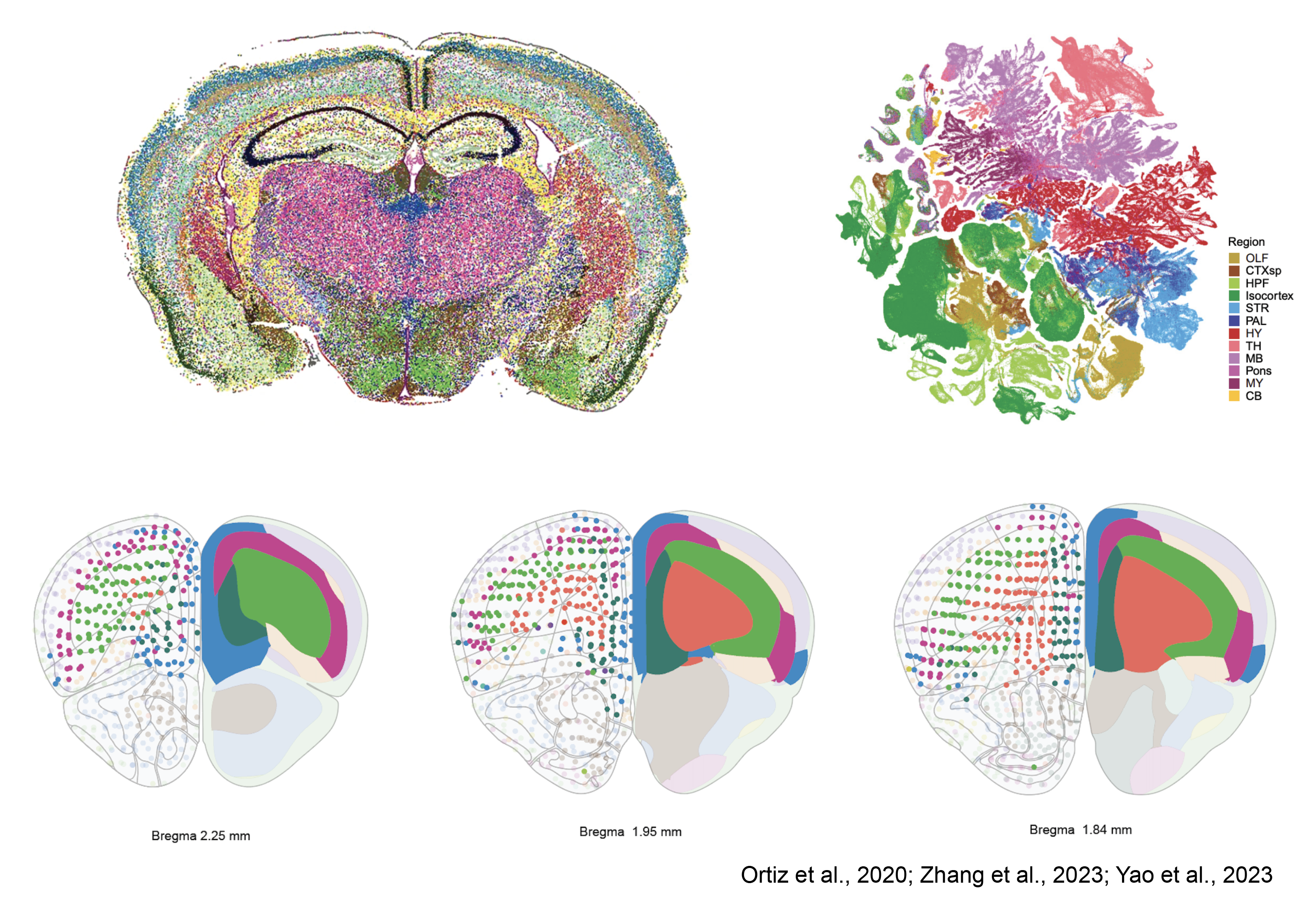
Mammalian brain regions embody three-dimensional architectures of neuronal assemblies established during development and evolution. Advancements in sequencing tools now enable us to discern the spatial patterns of gene expression profiles. In essence, brain regions and subregions are outlined solely based on their gene expression classification. This method unveils a laminar organization within the frontal cortex, aligning with what is traditionally recognized as the PFC. Thus, once again, the regions of the PFC identified by cytoarchitecture do not conform to the available gene expression maps. Despite being in the early stages of development, spatial transcriptomics and comprehensive genetic profiling of entire brain regions present a perplexing scenario. The lack of agreement between cytoarchitecture, connectivity and genetic identity remains to say the least, a puzzle that beckons further exploration.
Towards a functional map of the prefrontal cortex
Can functional properties of prefrontal neurons help us to understand the cellular logic of prefrontal operations? Can we use functional properties of neurons to make a map of brain regions? My current project in Pr. Carlén's laboratory is to use new methodological and analytical approaches to establish a functional map of the mouse prefrontal cortex. Here are some of the tools we use.
Research synthesis of mouse prefrontal functions
The prefrontal cortex has been ascribed many functions. in a recent perspective article, we have summarized the last decade of mouse PFC research. The data and methodology used to perform the functional research synthesis presented in the figure 3 of our perspective article: The mouse prefrontal cortex: Unity in diversity. The code used to generate the figures can be found on this github repository
Database
The database used to perform our research synthesis is available as a table. This table has the following columns: the year of the study, the short publication reference, the PFC region name used in the original publication, the inactivation method used, the stereotaxic triplet provided in the original article. The averaged coordinates used for the 3D plot into the Allen reference Atlas (ARA) CCFv3, the correction applied to the DV value to translate it into the ARA (see also below), the 3 scores; complexity index, sensory modality and task type, obtained for every publication, 7 logical values (in MOs, in ACA, in PL, in ILA, in ORBm, in ORBvl and in ORBl) indicating in which PFC region the study is found when plotted into the ARA CCFv3 with our method and the name of the Allen PFC region containing the stereotaxic triplet provided in the original publication.
The full database table can be downloaded here as an Excel or csv file.
3D visualisation
Here is an attempt of 3D visualisation of the database using plotly. Some improvements are still required. The Task type and Sensory Modality parameters are displayed as continuous variables although they are discrete variables. You can mouse over the dots in the scatter plot to see from which study is the data ONLY if you have already scrolled within the brain mesh (plotly feature). We hope to improve this 3D visualisation with time.
Limitations
The CCFv3 version of the Allen Atlas is a 10um voxel resolution space and our analysis using this Atlas has some limitations. We generated 3D meshes for the brain outline, for each PFC region (MOs, ACA, PL, ILA, ORBm, ORBvl and ORBl) and for the PFC sudivisions described in the section of our Perspective “Parcellations of the PFC” (dmPFC, vmPFC and vlPFC) by using the MATLAB “isosurface.m” function (see folder Brain_meshes). To detect in which Allen PFC region each publication was peformed we used the “inpolyhedron.m” function (Copyright © 2015, Sven Holcombe). The thickness of the borders in the actual version of the CCFv3 is around 30um (3 voxels). As a result some triplet of coordinates are allocated to 2 regions while performing our “inpolyhedron.m” detection procedure. We inspected manually each triplet of coordinates whithin its respective ARA plate (see folder AllenAtlasLocationCCFv3_individualstudies) and visually assesed for these ambiguous cases to which Allen PFC region they belong to. Our level of incertitude regarding PFC region assignment needs to be put into perspective because, as accurate as we can be as experimenters, the provided stereotaxic coordinates in every publication are only averaged values reflecting the mean location of the targetted brain structures. This experimental incertitude is by itself an important limitation of our analysis and needs to be taken into consideration.
Source
2015 Allen Institute for Brain Science. Allen Mouse Brain Atlas (2015) with region annotations (2017).
High-density electrophysiology
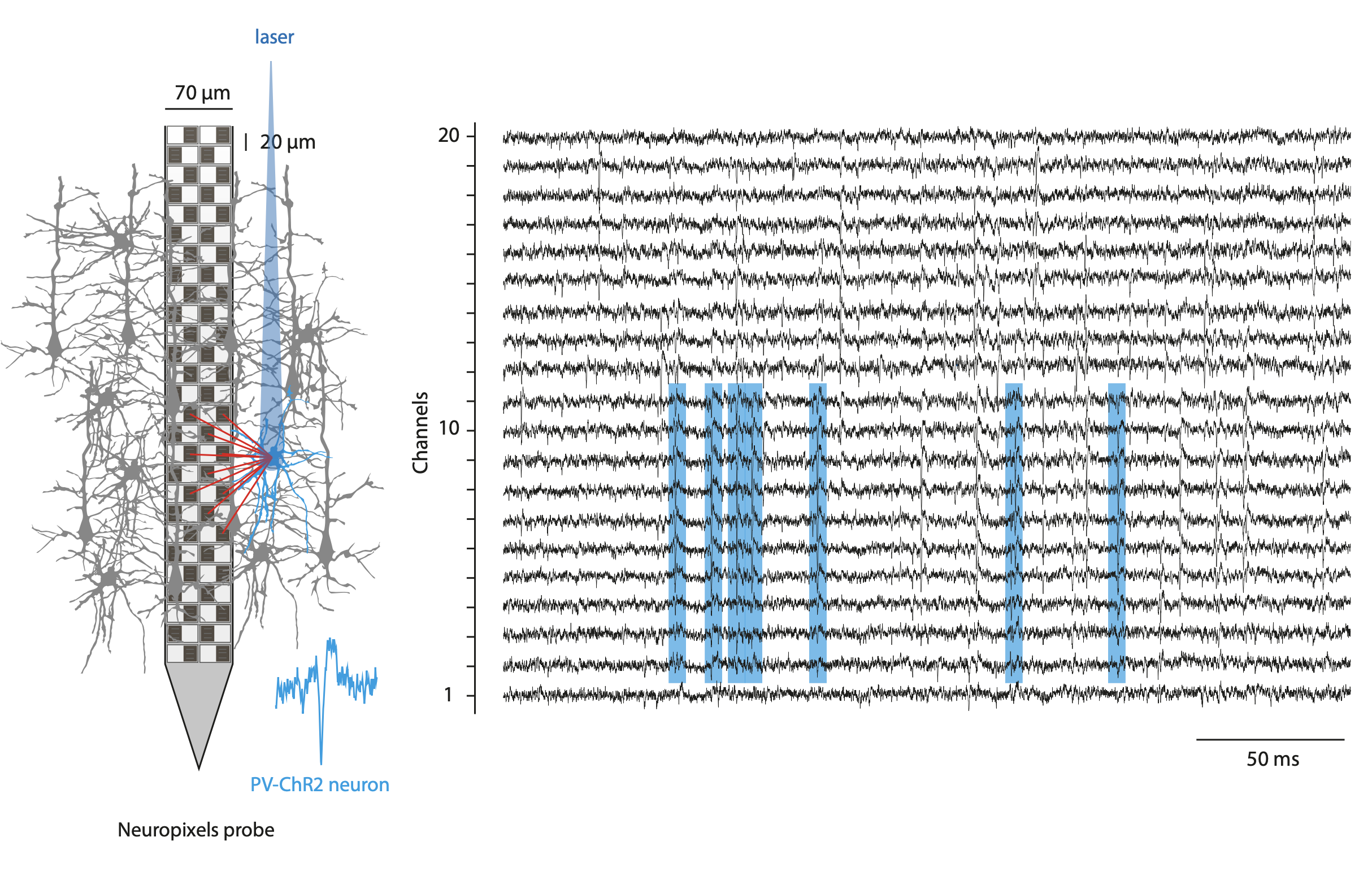
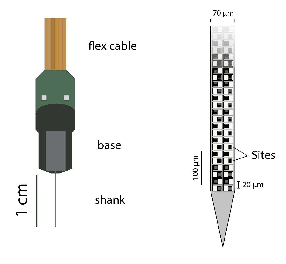
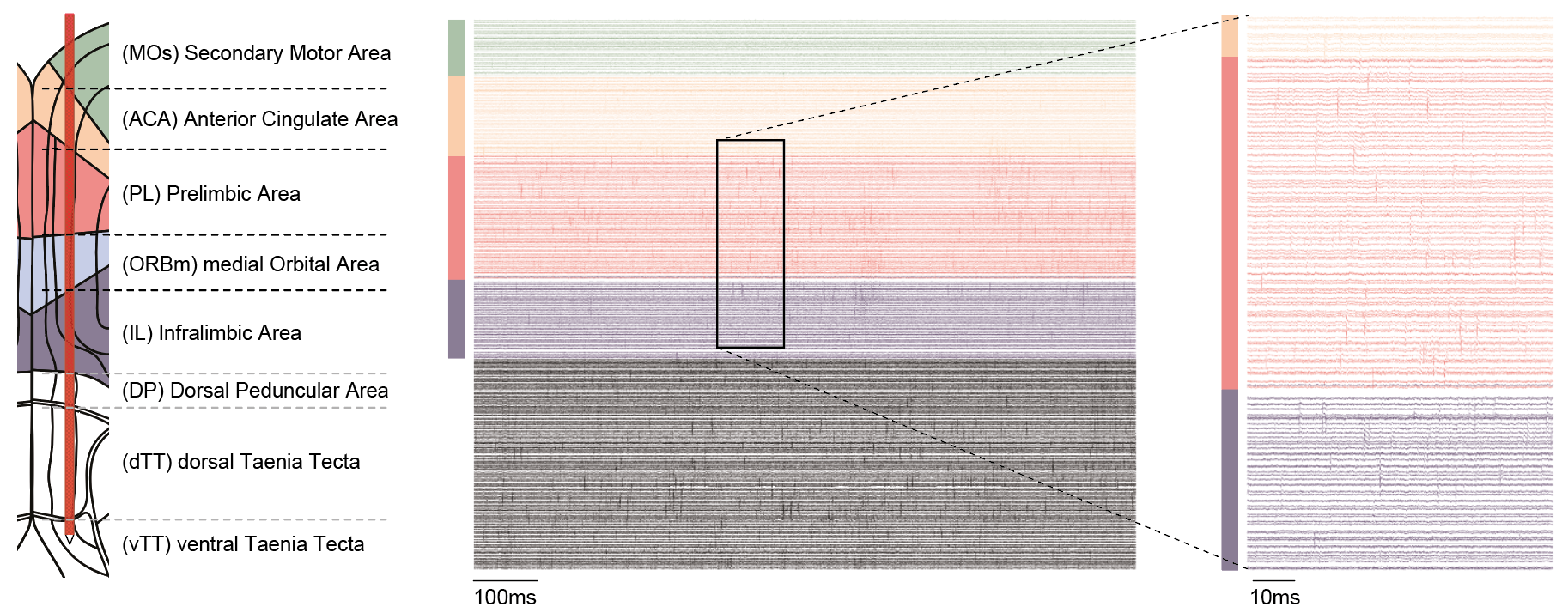
To record simultaneously the activity of hundreds of neurons from different prefrontal regions, I have been using the recently developped Neuropixels probes. These fully-integrated silicon CMOS digital neural probes (IMEC) allow to sample the extracellular neuronal activity simulataneously on 384 channels at a millisecond resolution. Combined with offline automated spike sorting algorithms, they offer for the first time the opportunity of recording units form many prefrontal regions in vivo. We use this system to gain knowlegde about the functional properties on the mouse prefrontal cortex regions while the mice engage in various behavioral tasks.
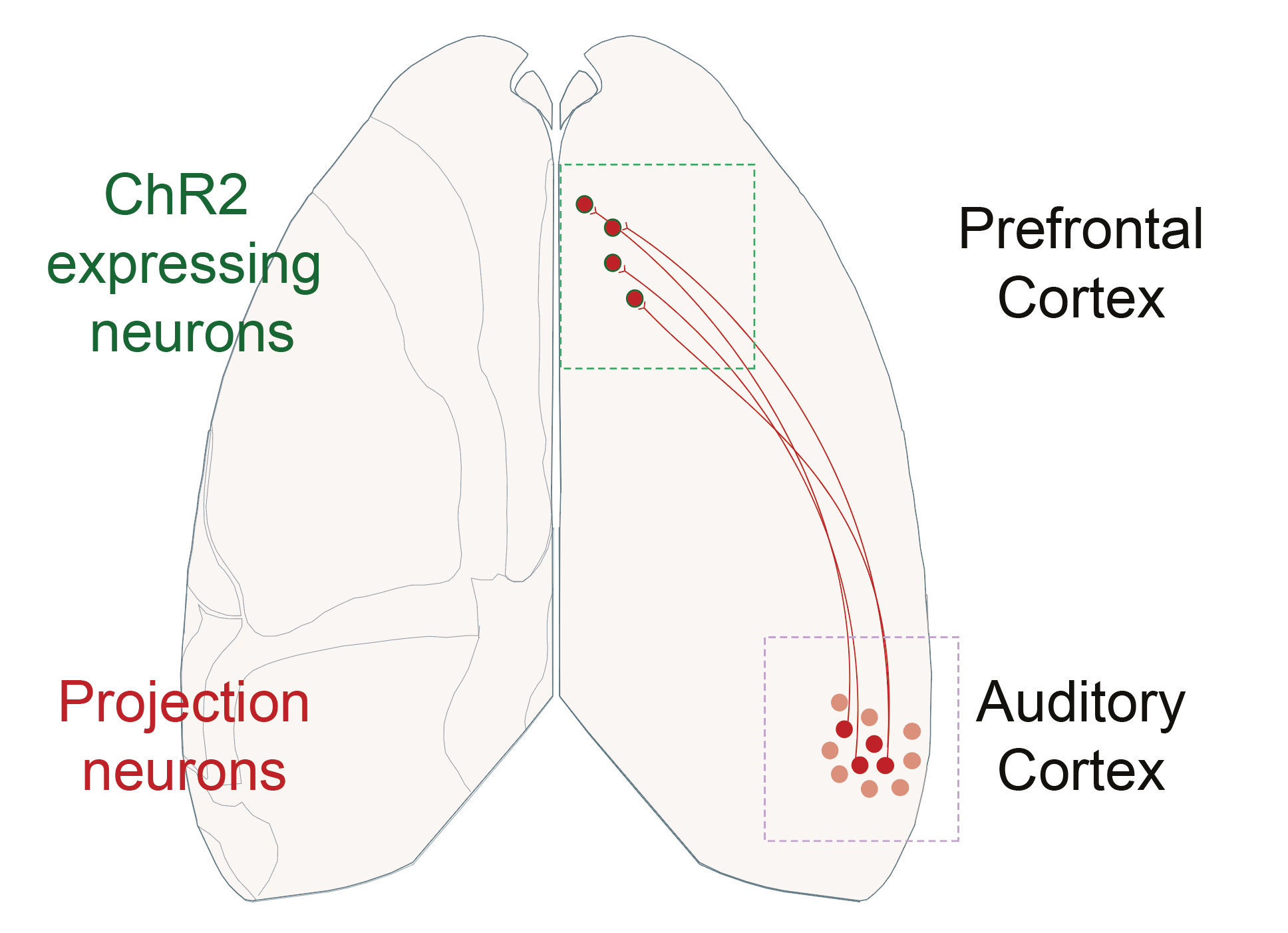
Optogenetic manipulations
As one of the classical technique utilized in neuroscience, I use optogenetics manipulations to interrogate the role of the prefrontal regions and cell types of interest. I have principaly used the optogenetic approach to photoinhibit cortical regions and to optotag neuronal populations of interest.